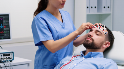
What Is an EMG Machine and How Does It Work?

An Emg Machine turns invisible muscle activity into a measurable electrical signal. When a person contracts a muscle, motor units produce tiny voltage changes. Surface electrodes detect these changes through the skin. Needle electrodes can measure activity closer to individual muscle fibers, when clinically appropriate.

Dr. Carlo J. De Luca, a widely recognized electromyography researcher, wrote, “The EMG signal is the electrical manifestation of the neuromuscular activation associated with muscle contraction.” This principle explains the machine’s basic role. The electrodes collect the signal. A differential amplifier strengthens it. Filters reduce interference from movement, nearby muscles, and electrical equipment. An analog-to-digital converter then sends the signal to software, where clinicians can view waveforms, amplitude, frequency, or timing.

The process sounds simple. It is not always simple.

A trained professional must place electrodes carefully. Skin preparation matters. Electrode distance matters. Even a relaxed hand can introduce noise through small, unnoticed movements. The displayed waveform is therefore not a perfect photograph of muscle function. It is an interpreted measurement.

This article will explain what an Emg Machine is, how its hardware works, and why signal quality affects clinical conclusions. It will also examine common applications, including diagnosing nerve disorders, assessing muscle recruitment, and supporting rehabilitation. These systems can provide valuable evidence, but they do not replace medical judgment. Results require patient history, physical examination, calibration checks, and appropriate professional interpretation. Small technical errors can change the story. That limitation deserves attention.
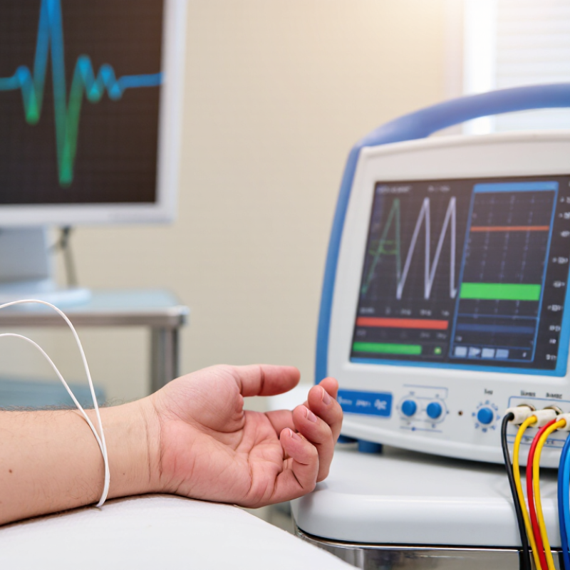
What Is an EMG Machine and How Does It Work?

What Is an EMG Machine?

An EMG machine is a medical system that records electrical activity in muscles and nerves. EMG stands for electromyography. The device uses small electrodes to detect signals produced when muscle fibers contract. A computer then displays these signals as waves, sounds, or numerical readings.

A clinician may place surface electrodes on the skin over a selected muscle. These flat sensors can capture activity during rest and movement. In some examinations, a very fine needle electrode enters the muscle briefly. The needle records activity from a smaller area, which can provide more specific information. The machine amplifies these tiny signals because they are usually too weak to observe directly.

During a test, the patient may relax, tighten a muscle, or repeat a controlled movement. I have found that even a simple action, such as lifting a finger, can create a clear change on the screen. The pattern may help a trained specialist assess muscle function, nerve communication, or possible nerve injury. However, the machine does not diagnose a condition by itself. Medical history, physical examination, and other test results still matter.

The signal is not always clean. Movement, cold skin, loose electrodes, or nearby electrical devices can create noise. Results also depend on electrode placement and the examiner’s technique. That part is easy to underestimate. A careful clinician checks the recording quality before interpreting the waves.

The Main Components of an EMG System

What Is an EMG Machine and How Does It Work?

The Main Components of an EMG System

An EMG system records the electrical activity produced by muscles. Its main sensing parts are surface electrodes or fine needle electrodes. Surface electrodes rest on cleaned skin, while needle electrodes measure activity inside a specific muscle. A reference electrode helps reduce unwanted electrical interference. Accurate placement matters greatly. A few millimeters can change the recorded signal.

The electrode cables connect to a preamplifier. This stage receives very small voltage changes and strengthens them without adding excessive noise. The system then uses filters to reduce interference from movement, power lines, and nearby equipment. Filtering must be carefully controlled. Too much filtering may hide clinically useful details.

A computer interface converts the conditioned analog signal into digital data. Specialized software displays waveforms, audio signals, amplitude, duration, and recruitment patterns. Some systems also include a stimulator for nerve conduction testing, which uses controlled electrical pulses. During a practical examination, the operator checks the equipment, prepares the skin, and observes the patient’s movement and comfort. Small artifacts can still appear, even with careful preparation. That imperfection requires judgment, not automatic trust in the screen. A trained clinician interprets the findings alongside symptoms, physical examination, and medical history. The machine measures signals; it does not independently diagnose disease.

What Is an EMG Machine and How Does It Work? - The Main Components of an EMG System

System Component Primary Function What It Measures or Processes Typical Technical Characteristics Role in the Examination
Surface Electrodes Detect electrical activity from muscles through the skin. Summed activity from multiple nearby motor units. Usually disposable adhesive electrodes made with conductive materials; commonly used for nerve conduction studies and surface recordings. Placed over a muscle or nerve pathway to record responses without inserting a needle.
Needle Electrode Records electrical activity directly inside or near a selected muscle. Motor-unit action potentials, spontaneous activity, and changes during muscle contraction. Fine sterile needle electrode with a small recording surface; used only by trained clinical personnel. Provides detailed information about muscle fibers, motor units, and possible denervation or reinnervation.
Reference Electrode Provides a stable electrical reference for differential recording. Electrical potential at a relatively inactive location near the recording site. Positioned away from the active recording area and secured to the skin. Helps the system distinguish the desired biological signal from common electrical interference.
Ground Electrode Reduces electrical interference and improves recording stability. Provides a common reference point for rejecting environmental noise. Attached to the patient, typically between the stimulus or recording site and the amplifier. Supports safer, cleaner measurements but does not replace electrical safety protections.
Electrical Stimulator Delivers a controlled electrical pulse during a nerve conduction study. Nerve response to a known stimulus intensity, duration, and location. Adjustable pulse amplitude, duration, polarity, and repetition rate; output is controlled by the examination protocol. Activates a peripheral nerve so latency, amplitude, and conduction velocity can be calculated.
Preamplifier Receives the very small signal from the electrodes before further processing. Low-level bioelectric signals, often affected by motion and electromagnetic noise. High input impedance and differential inputs help reduce signal loss and common-mode interference. Provides the initial signal conditioning needed for accurate EMG and nerve conduction recordings.
Main Amplifier Increases the signal to a level suitable for display, storage, and analysis. Amplified muscle and nerve electrical activity. Adjustable gain, differential amplification, and common-mode rejection; settings vary by test type. Allows low-amplitude physiological signals to be viewed and analyzed without excessive distortion.
Filters Reduce unwanted frequencies and improve signal readability. Frequency content of the recorded EMG or evoked response. High-pass, low-pass, and sometimes notch filtering; filter settings must be selected carefully to avoid removing clinically useful information. Limits artifacts such as motion, electrical mains interference, and high-frequency noise.
Analog-to-Digital Converter Converts the conditioned analog signal into digital data. Voltage changes sampled over time. Sampling rate and resolution determine how accurately rapid waveform changes and small amplitude differences are represented. Enables digital display, measurement, storage, waveform review, and computer-based analysis.
Acquisition and Analysis Software Controls test protocols and analyzes recorded signals. Latency, amplitude, duration, conduction velocity, waveform morphology, and recruitment patterns. Provides configurable examination settings, waveform measurement tools, annotations, and report generation. Helps the examiner document findings and compare results with clinical reference values.
Display and Audio Output Presents electrical activity visually and, in many systems, acoustically. Waveforms, numerical measurements, and audible changes in motor-unit activity. Real-time traces with adjustable time base, amplitude scale, sweep speed, and audio volume. Allows the examiner to observe waveform shape and listen for changes during voluntary contraction.
Patient and Electrode Cables Connect electrodes and stimulators to the recording unit. Biopotential signals and stimulation pulses. Shielded, color-coded, low-noise cables with connectors designed for secure attachment. Maintains signal integrity and supports correct electrode placement throughout the test.
Electrical Isolation and Safety Circuits Protect the patient and operator from hazardous leakage currents. Electrical connection between the patient-connected circuitry and external power sources. Uses isolated patient circuits, protective grounding, current limitation, and safety monitoring according to medical-device requirements. Provides essential protection during recording and electrical nerve stimulation.

An EMG system records electrical signals produced by muscles and peripheral nerves. Needle EMG primarily evaluates activity within a muscle, while nerve conduction studies use surface electrodes and controlled electrical stimulation to assess nerve response.

How an EMG Machine Records Muscle Activity

What Is an EMG Machine and How Does It Work?

An EMG machine records the electrical activity produced by muscles. When a muscle contracts, nerve signals activate tiny muscle fibers. These fibers create brief electrical impulses, called motor unit action potentials. The machine detects and displays these signals for a trained clinician.

How an EMG Machine Records Muscle Activity

For surface EMG, small adhesive electrodes are placed on clean skin above a muscle. The electrodes sense voltage changes while the person rests, tightens the muscle, or performs a guided movement. The machine amplifies these low-level signals and sends them to a monitor. A waveform may rise sharply during contraction, then fall as the muscle relaxes. Some systems also produce sound, allowing the clinician to hear changes in activity.

Needle EMG uses a fine electrode inserted into the muscle. It can capture activity from a smaller area than surface electrodes. The clinician may ask the patient to relax, make a gentle contraction, and then increase effort. The screen can reveal the timing, size, and pattern of electrical signals. These details may support assessment of nerve or muscle disorders, but an EMG is not a perfect window into muscle health. Movement, sweat, electrode placement, and nearby muscles can distort recordings. I have found that even a small shift in an electrode may change the waveform. Results therefore need clinical experience, patient history, and other tests for proper interpretation.

The Step-by-Step EMG Testing Process

What Is an EMG Machine and How Does It Work?

The Step-by-Step EMG Testing Process

An EMG machine records electrical signals from muscles and nerves. A trained clinician usually performs the test in a quiet examination room. Before testing, they explain each step and review symptoms, medicines, and relevant medical history. The patient may sit or lie down with the limb supported.

The clinician cleans the skin to improve signal quality. Small surface electrodes are placed over selected muscles. The patient then relaxes, gently tightens the muscle, and gradually increases effort. The machine displays changing waveforms on a monitor. These patterns can show how muscles respond at rest and during movement.

Some examinations also use a fine needle electrode. The clinician inserts it into specific muscles and records activity during rest and contraction. A brief electrical pulse may test nerve conduction in nearby areas. It can feel sharp, awkward, or mildly uncomfortable. Most people tolerate it well.

The clinician compares findings with the physical examination and symptom history. One waveform never tells the whole story. Temperature, muscle tension, electrode placement, and patient movement can affect results. A rushed explanation can also increase anxiety. Results should therefore be interpreted by a qualified healthcare professional, not by the machine alone. The report may help assess nerve compression, muscle disorders, or nerve damage, but further evaluation is sometimes needed.

How Doctors Interpret EMG Results

An EMG machine records electrical activity from muscles and nerves. Doctors usually assess two related tests: nerve conduction studies and needle EMG. Small electrodes deliver gentle electrical impulses through the skin. The equipment measures latency, amplitude, and conduction speed. A fine needle electrode then records muscle activity at rest and during contraction.

Doctors do not interpret one number alone. They compare each result with reference ranges, the patient’s symptoms, and the physical examination. Slower conduction may suggest nerve compression, while reduced amplitude can indicate axonal nerve damage. Abnormal spontaneous activity, such as fibrillation potentials, may show muscle denervation. During movement, the size and recruitment of motor units provide additional clues.

Pattern matters most.

For example, findings across several nerves may support a generalized neuropathy. Abnormal results limited to one nerve may fit local entrapment. However, temperature, electrode placement, age, medications, and technical factors can influence measurements. A borderline result can invite overdiagnosis. That deserves caution.

A normal EMG also has limits.

It may not detect very early disease, intermittent symptoms, or small-fiber disorders. Doctors therefore combine the report with medical history and examination findings. The test can feel uncomfortable, but most discomfort is brief. Patients should report pain, weakness, numbness, and symptom timing accurately. Interpretation is clinical judgment, not an automatic machine verdict.

Close

Raise your Query

Hi! Simply click below and type your query.

Our experts will reply you very soon.

WhatsApp Us